Friday 30 September 2016

PCP Pneumonia

RV positive patient since 16 yrs with recent CD 4 of 109 came with :

Fever with chills since 2mts, dry cough since 1 month, reduced appetite  since 1 month and weight loss of 12 kg over past one month. 


The chest x-ray findings were thought to be related to Koch's and he was started on AKT....


There are coarse reticular opacities in bilateral para hilar and para-cardiac region.Few small cysts were seen in the mid zones.

 Lecopenia was seen ( 1782 /cumm)

Bronchoscopy guided bronchial aspirates revealed PCP organisms. 

Monday 29 August 2016

Haglund Syndrome

Posterior heel pain


Triad of HS on plain radiograph-

  1. Achillis Tendinopathy( thick AT at its insertion with enthesophytes)  
  2. Retro-calcaneal bursitis ( Obliteration of Kager's fat pad)
  3. Haglund deformity ( bony bump at the posterior superior aspect of the calcaneus)

Friday 3 June 2016

Ankle Joint osteoarthritis /Talar Osteochondritis dessicans

50 yrs female with ankle Pain since few months. She is known case of OA Knee.

Add caption
Subarticular sclerosis along dome of talus on lateral aspect( red arrow)  and subarticular lytic area in the medial aspect.
Irreularity along superior aspect of the neck of talus.

Possibility of osteoarthritis of ankle joint is more likely and is probably secondary to OA Knee with produces undue strain on the ankle joint while walking.


Normal Ankle Joint of another patient for comparison


Etiology of Ankle OA:

  • Post traumatic ( most common)- Traumatic ankle injuries that may result in OA include fractures of the malleoli, tibial plafond, talus, isolated osteochondral damage of the talar dome, and ankle ligament injury
  • Primary OA, ie, OA with no history of trauma or any kind of secondary or infectious OA, in the ankle is rare.
  • Secondary to rheumatoid arthritis and rarely due to hemochromatosis or hemophilia.
  • Varus alignment may be a predisposing factor.


Ankle has high resistance cartilage cover and limited range of motions as compared to knee joint so risk of primary OA is less.


Friday 27 May 2016

Haglund Syndrome with Normal Os Peroneum

A female patient with pain in the posterior aspect of the heel-

Os peroneum is normally located lateral to the cuboid. Accesory Navicular is also noted

Thickening of Achilis tendon at its insertion with enthesophyte and adjacent soft tissue swelling. There is mild  loss of normal retro-calcaneal luceny s/o retro-calcaneal bursitis
Haglund Syndrome:
It is said to be present in cases of patients with heel pain if following signs are present-

  1. Achillis insertional site enthesophytes.
  2. Insertional Achillis tendinitis,
  3. Retro-calcaneal( Kager's fat) and Retro-achillis bursitis.
  4. Bone marrow edema in calcaneal tuberosity( MRI)
Predisposing factors: Hind foot varus and pes cavus. 
Those wearing low back shoes are prone for HS-

Os Peroneum:
Located adjacent to calcaneo-cuboid joint( lateral radiograph- 7mm proximal -8mm distal to CC joint, Oblique radiograph- .9mm proximal and 8mm distal to CC jt)
It lies within the peroneus longus tendon.
It can be uni-partite/bi-partite or multi-partite( margins have to be well defined and corticated with minimal i.e. less than 2mm inter-fragment distance).

Fracture of Os Peroneum- os peroneum fragment separation of 6 mm or more or displacement of the proximal Os peroneum fragment by 10 mm or more proximal to the calcaneocuboid joint on a lateral radiograph or by 20 mm or more proximal to the calcaneocuboid joint on an oblique radiograph. It is associated with full-thickness tear of the peroneus longus tendon.

Heel Pain Causes:

The most common causes of heel pain are plantar fasciitis (bottom of the heel) and Achilles tendinitis (back of the heel).
Differential Diagnosis Considerations:

Arthritic -Gout, rheumatoid arthritis, seronegative arthropathy; primary or secondary
osteoarthritis
Infectious -Diabetic ulcer, osteomyelitis, plantar warts
Mechanical 
Plantar—
plantar fasciitis, heel spur, calcaneal stress fracture, medial or lateral
plantar nerve entrapment, heel pad syndrome, foreign body granulomatosis
Posterior—
Achilles tendinopathy, Haglund deformity, retrocalcaneal bursitis, tarsal
coalition, accessory muscles( accesory soleus muscle in posteromedial heel pain), Sever disease (pediatrics)
Neuropathic- Lumbar radiculopathy, nerve entrapment, neuroma, tarsal tunnel syndrome
Trauma
Tumor (rare) -Ewing sarcoma, neuroma
Vascular (rare)


Saturday 16 April 2016

Inflammatory Bowel disease: Crohn's

A 40 yrs female with history of loose non bloody watery stools, weight loss and odynophagia since few months-


The Barium Meal Follow Through Spot compression images reveal eccentric narrowing of the distal small bowel loop with anti-mesenteric pseudosacculations.  The distal ileum stands distinct due to mesenteric fibro-fatty proliferation displacing it from rest of the small bowel. Cecum also shows cobble stone appearance. 


Large bowel colonsocopy upto hepatic flxure reveals serpiginious ulcers with spared normal mucosa giving cobble stone appearance relatively sparing rectum.


Monday 11 April 2016

Small bowel strictures at two distinct sites involving ileum as detected on Small Bowel Enteroclysis:


There is prestenotic dialation. The strictures are short segment.
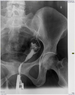
Traumatic rupture of Bulbar Urethra. Pelvic bone fractures with peri-prostatic hematoma displacing the bladder upwards:


Twisting injury to Foot : Avulsion injury to base of 5th Metatarsal


The fractures at the base of the 5th metatarsal are transversely oriented to the shaft of metatarsal.
Unfused apophysis is laterally located and is longitudinally oriented ( It is seen on radiograph  at 10 to 12 yrs in girls and boys respectively and fuses within next 2-4yrs).

Another case:

Here the line at the base of 5th metatarsal is transversely oriented and only involves the tip so it is a avulsion fracture.

This is most common avulsion injury of foot and most common type of injury ( 90%) at the base of the 5th metatarsal.  It is caused due to forceful plantar inversion in flexed foot of postion with avulsion of the insertion of peroneus brevis. It may or may not extend into the metatarso-cuboid joint.

Accesory ossicle just at the base of 5th metatarsal is called Os Vesalianum( very rare) while lateral to the cuboid is called Os Peroneum are other differentials but they are well corticated seasmoids.

Islein Disease: is apophysitis of the base of 5th metatarsal due to repetitive traction by peroneus brevius usually in young persons engaged in the sports activities. There is irregularity of the apophysis or bony specks in this region.

Thus Most common fracture at the base of 5th metetarsal is Avulsion injury , f/b Jones f/b Stress fracture.

Saturday 9 April 2016

A young male came with complains of acute retention of urine and supra-pubic pain. He gives recent history of a obstructive right ureteric calculus.

.


RADIOGRAPH ABDOMEN (KUB) – SUPINE A. P. VIEW

Poor bowel preparation.
Small 6mm and 4mm radiopacities in the region of symphysis pubis probably urethral calculus. Kindly correlate with USG KUB.

No other abnormal calculus density is noted in the region of urinary tract.

Impacted Urethral Stone.

In a KUB Radiograph include the symphisis pubis region so as not to miss out this.

Wednesday 30 March 2016

Os Trigonum


Can be mistaken for avulsion fracture of lateral tubercle of talus( Shepherd's fracture) or Fracture of steida process. Normally it is well corticated.

It may be the the source of Os trigonum syndrome or posterior ankle impingement syndrome in people performing repetitive plantar flexion activities. Posterior recess synovitis of the tibio-talar and posterior subtalar joint can occur due to impingement.  Flexor halluics tendon is located medial to the Os-trigonum and can get injured in the process.

Os trigonum is a secondary ossification center in the posterolateral aspect of the talus, which is present in approximately 5%-15% of “normal” feet. The ossification occurs between 7 and 13 years of age, and, within one year, the Stieda’s process is formed; however, it can remain as a separate ossicle in 7%- 14% of patients, usually bilaterally.

It is worth noting that the following individuals are more prone to develop Os Trigonum Syndrome: athletes of sports involving kicking; ballet dancers who assume the en-pointe and demi-pointe positions; and workers who use pedals, such as drivers and seamstresses.
 On physical examination, palpation of the posterior portion of the ankle joint, as well as
the passive maximum plantar flexion maneuver, causes pain.


Child with cough and fever since 15 days:


Right paratracheal and left hilar nodes favour mediastinal Tb.
Lungs are clear.

Lymph node Tb constitutes 20-40% of extra-pulmonary Tb. It is more common in children and women than other forms of extra-pulmonary Tb. It is more common in Asians and Pacific islanders. In developing and underdeveloped countries , it continues to be caused by M.Tuberculosis and atypical mycobacteria are seldomly isolated.

Commonly involved superficial lymph nodes are-

  • Posterior and anterior cervical chain.
  • Submandibular, peri-auricular, inguinal and axillary groups.
  • Intra-throacic ( hilar-paratracheal and mediastinal in decreasing order of frequency)
  • Abdominal nodes.


Frequency of associated pulmonary involvement is 5-62%.

Most patients can be managed medically and surgical intervention is rarely required

? Odontoid fracture

History of Trauma:

Cervical spine- Flexion and Extension View:


Atlanto Occipital assimilation and odontoid tip fracture. Confirmed on CT

Tuesday 22 March 2016

BIRADS 5 lesion

A middle aged female with pain and lump in left breast-

Right breast:


Left Breast:


Upper -outer quadrant spiculated mass in left breast ( BIRADS 5)


Ref:

http://www.radiologyassistant.nl/en/p53b4082c92130/bi-rads-for-mammography-and-ultrasound-2013.html

Post Traumatic Osteolysis of lateral end of right clavicle


Bilateral erosions


  • hyperparathyroidism

subchondral bony resorption; usually symmetric with osteopenia, abnormal trabecular pattern
the acromion is normal, but the sternoclavicular joint may be affected

  • rheumatoid arthritis

bilateral or unilateral changes with soft tissue swelling, subchondral osteoporosis and erosion of the outer third of the clavicle
acromial erosions may occur later in the disease process

  • scleroderma
  • cleidocranial dysostosis
  • pyknodysostosis (rare)

Unilateral erosion


  • post-traumatic osteolysis
  • myeloma
  • metastases
  • osteomyelitis

Cardiac Calcification's

1) Known case of Rheumatic Valvular heart disease and coronary artery disease-
Dense Mitral Annulus calcification
Approximate location-
Brown ring- Mitral valve
Yellow ring- aortic valve
Green ring-pulomnary valve
Purple ring- tricuspid valve
Dark red ring- aortic knuckle
Blue line- pericardial calcification


This case demonstrates calcification of the mitral valve annulus (not to be confused with mitral valve leaflet calcification which is the result of, and can cause, mitral valve disease).
Coarse calcification is seen in the expected location of the mitral valve, to the left of midline.  It is associated with conduction defects and coronary artery disease.
Other causes-
  • Metastatic calcification in the form of myocardial calcinosis  is an entity need to be considered in patients with bone disease, hypercalcemia, hyperphosphatemia, renal disease or those on chronic dialysis. Complications arising from cardiac calcification include valvular dysfunction, complex atrial and ventricular arrhythmias, coronary events and sudden cardiac death.
  • Calcified Peri-cardial cyst
  • Calcified hydatid cyst of heart
  • Intra-cardiac calcified aneurysm
  • calcified old myocardial infarct
  • Rheumatic valvular disease with calcification of the valvular leaflets
  • Atrial appendage calcification ( also in RVHD and myocardial calcinosis)


Tuesday 9 February 2016

Uterine anomalies

1) 

There is single endometrial cavity.
It is elongated and banana shaped.
It is deviated to left side.
It has tapered end.
Single cervix is seen.
Single left Fallopian tube is patent.
No contrast spill on right side.

These features indicate Left Unicornuate uterus with patent left fallopain tube.
There is no e/o of a communicating right sided rudimentory horn, however a non-communicating one can be made out on MRI/ 3D USG.

Sono HSG is useful modality in demonstrating uterine anomalies.

These patients are at risk of spontanoeus abortions, preterm delivery, abnormal fetal position and IUGR

Types of Mullerian duct anomalies-


Friday 8 January 2016

Type II Salter Harris

A child with history of trauma-


Type II SH fracture at distal end of radius.Ulnar styloid fracture.


Cervical Tb

22 yrs female -


There is prevertebral soft tissue swelling with erosion of anterior cortex of C2 vertebral body.
Features favour Tuberculosis.


Atlanto-axial Instability noted in flexion-extension views

Pathological fracture  probably due to duerlying NOF(Non-Ossifying Fibroma)

RV positive patient with Tuberculosis cavitatory lesions